Mostrar mensagens com a etiqueta LESÕES NO DESPORTO. Mostrar todas as mensagens

domingo, 29 de novembro de 2009

ARTIGO DO MÊS - 28/2009

Anterior Cruciate Ligament Deficiency Causes Brain Plasticity
Kapreli, E;  Athanasopoulos, S.; Gliatis, J.; Papathanasiou, M. ; Peeters, R.;  Strimpakos, N.;  Hecke, P.V.;  Gouliamos, A.; Sunaert, S.
Am J Sports Med 2009 37: 2419


Background: The mechanoreceptors located in anterior cruciate ligament (ACL) constitute an afferent source of information toward the central nervous system. It has been proposed that ACL deficiency causes a disturbance in neuromuscular control, affects central programs and consequently the motor response resulting in serious dysfunction of the injured limb.


Purpose: The objective of this study was to investigate whether chronic anterior cruciate ligament injury causes plastic changes in brain activation patterns.


Study Design: Case control study; Level of evidence, 3.


Methods: Seventeen right leg–dominant male participants with chronic anterior cruciate ligament deficiency and 18 matched healthy male participants with no special sport or habitual physical activity participated in this study. Patient selection criteria comprised a complete right unilateral anterior cruciate ligament rupture ≥6 months before testing. Brain activation was examined by using functional magnetic resonance imaging technique (1.5-T scanner).


Results: Results show that patients with anterior cruciate ligament deficiency had diminished activation in several sensorimotor cortical areas and increased activation in 3 areas compared with controls: presupplementary motor area, posterior secondary somatosensory area, and posterior inferior temporal gyrus.


Conclusion: The current study reveals that anterior cruciate ligament deficiency can cause reorganization of the central nervous system, suggesting that such an injury might be regarded as a neurophysiologic dysfunction, not a simple peripheral musculoskeletal injury. This evidence could explain clinical symptoms that accompany this type of injury and lead to severe dysfunction.Understanding the pattern of brain activation after a peripheral joint injury such as anterior cruciate ligament injury lead to new standards in rehabilitation and motor control learning with a wide application in a number of clinical and research areas (eg,surgical procedures, patient re-education, athletic training, etc).


Keywords: anterior cruciate ligament (ACL) deficiency; noncopers; central nervous system; reorganization; plasticity; functional magnetic resonance imaging (fMRI)


(...) rehabilitation should focus on CNS reeducation rather than optimizing only the peripheral neuromuscular function. Therapists should take advantage of the adaptive abilities of the CNS, identifying the goals, providing the tools, and allowing the CNS to find a solution.
(...) rehabilitation  should be based on procedures that stimulate the learning process and CNS reprogramming

domingo, 15 de novembro de 2009

ARTIGO DO MÊS - 27/09

A Systematic Review of Anterior Cruciate Ligament Reconstruction with Autograft Compared with Allograft
Carey,J.; Dunn, W.R.; MPH, Dahm, D.L.; Zeger, S.L.; and Spindler, K.P.
J. Bone Joint Surg Am. 2009;91:2242-50

Background: Anterior cruciate ligament reconstruction can be performed with use of either autograft or allograft tissue. It is currently unclear if the outcomes of these two methods differ significantly. This systematic review and meta-analysis investigated whether the short-term clinical outcomes of anterior cruciate reconstruction with allograft were significantly different from those with autograft.

Methods: A computerized search of the electronic databases MEDLINE and EMBASE was conducted. Only therapeutic studies with a prospective or retrospective comparative design were considered for inclusion in the present investigation. Two reviewers independently assessed the methodological quality and extracted relevant data from each included study. If a study failed the qualitative assessment and statistical tests of homogeneity, it was excluded from the meta-analysis. Furthermore, a study was withdrawn fromthe meta-analysis of a particular outcome if that outcome was not studied or was not reported adequately. A Mantel-Haenszel analysis utilizing a random-effects model allowed for pooling of results according to graft source while accounting for the number of subjects in individual studies.

Results: Nine studies were determined to be appropriate for the systematic review. Eight studies compared bone-patellar tendon-bone grafts, and one study compared quadruple-stranded hamstring grafts. Five studies were prospective comparative studies, and four were retrospective comparative studies. One study, which investigated allografts that underwent a unique sterilization process, demonstrated an allograft failure rate of 45% (thirty-eight of eighty-five). That study failed the qualitative assessment and statistical tests of homogeneity and consequently was excluded from the meta-analysis. When the outcomes from the remaining studies were pooled according to graft source, the meta-analyses of the Lysholm score, instrumented laxity measurements, and the clinical failure rate estimated mean differences and odds ratios that were not significant. These findings were robust during the sensitivity analysis, which varied the included studies or variables on the basis of graft type, instrumented laxity cut-off value, secondary sterilization technique, duration of follow-up, mean patient age, and study methodology.

Conclusions: In general, the short-term clinical outcomes of anterior cruciate reconstruction with allograft were not significantly different from those with autograft. However, it is important to note that none of these nonrandomized studies stratified outcomes according to age or utilized multivariable modeling to mathematically control for age (or any other possible confounder, such as activity level, that is not equally distributed in the two treatment groups). Understanding these limitations of the best available evidence, the surgeon may incorporate the results of the present systematic review into the informed-consent and shared-decision-making process in order to individualize optimum patient care.

Level of Evidence: Therapeutic Level III.

sábado, 7 de novembro de 2009

ARTIGO DO MÊS - 26/09

The Ability of 4 Single-Limb Hopping Tests to Detect Functional Performance Deficits in Individuals With Functional Ankle Instability

Erin Caffrey, Carrie L. Docherty, John Schrader, Joanne Klossner
DOI: 10.2519/jospt.2009.3042 in JOSPT NOVEMBER 2009, Volume 39, No. 11


STUDY DESIGN: Experimental laboratory testing using a cross-sectional design.

 OBJECTIVES: To determine if functional performance deficits are present in individuals with functional ankle instability (FAI) in 4 single-limb hopping tests, including figure-of-8 hop, side hop, 6-meter crossover hop, and square hop.

BACKGROUND: Conflicting results exist regarding the presence of functional deficits in individuals with FAI. It is important to evaluate whether functional performance deficits are present in this population, as well as if subjective feelings of giving way can assist in identifying these deficits.

METHODS: Sixty college students volunteered for this study. Thirty participants with unilateral ankle instability were placed in the FAI group and 30 participants with no history of ankle injuries were placed in the control group. The FAI group was subsequently further divided to indicate those that reported giving way during the functional test (FAI-GW) and those that did not (FAI-NGW). Time to complete each test was recorded and the mean of 3 trials for each test were used for statistical analysis. To identify performance differences, we used 4 mixed-design 2-way (side-by-group) ANOVAs, 1 for each hop test. A Tukey post hoc test was completed on all significant findings.

RESULTS: We identified a significant side-by-group interaction for all 4 functional performance tests (P<.05). Specifically, for each functional performance test, the FAI limb performed significantly worse than the contralateral uninjured limb in the FAI-GW group. Additionally, the FAI limb in the FAI-GW group performed worse than the FAI limb in the FAI-NGW group, and the matched limb in the control group in 3 of the 4 functional performance tests.

CONCLUSION: We found that functional performance deficits were present in participants with FAI who also experienced instability during the test. This difference was identified when comparing the FAI limb to the contralateral uninjured limb as well as control participants. However, the performance deficits identified in this study were relatively small. Future research in this area is needed to further evaluate the clinical meaningfulness of these findings. Finally, we found that limb dominance did not affect performance.

 
KEY WORDS: 6-meter crossover hop, agility, figure-of-8 hop, side hop, square hop


The authors determine if functional performance deficits are present in individuals with functional ankle instability (FAI) in 4 single-limb hopping tests, including figure-of-8 hop, side hop, 6-meter crossover hop, and square hop.

Comentário: Os testes de desempenho funcional representativos de cada segmento funcional e da sua coordenação/interacção com os segmentos adjacentes envolvendo os mecanismos neuromotores que os controlam podem ser variáveis a integrar quer na avaliação quer na reeducação/normalização da função.

terça-feira, 1 de setembro de 2009

LESÕES NO TÉNIS - CASOS REAIS (6)


RAFAEL NADAL após 2 meses de paragem regressou à competição nos Torneios Masters que antecedem o US Open 2009.
Para já parece estar recuperado, mas o teste principal vai ser no US Open onde os jogos são discutidos à melhor de 5 sets e em piso rápido.
Face ao histórico destas lesões - Tendinopatias crónicas - comentadas ao longo deste blog, os primeiros indicadores são positivos o que confirma a necessidade de repouso que Rafa foi obrigado associado aos tratamentos adequados que a equipa clinica que o acompanha realizou.
No entanto sugere-se moderação na participação dos principais Torneios com períodos de recuperação activa.
Faço votos para que o grande campeão espanhol volte em pleno.

Raul Oliveira, Fisioterapeuta
R´Equilibri_us - Gabinete de Fisioterapia
Av. D. João I, nº 8, Oeiras
309 984 508 /917231718/ 917776556
Faculdade de Motricidade Humana

sexta-feira, 26 de junho de 2009

LOMBALGIAS NOS ATLETAS (1)


A lombalgia ou dor lombar constitui uma das queixas mais frequentes dos jovens atletas sobretudo em determinadas modalidades desportivas.

Actualmente registamos 2 realidades distintas :

a) com o aumento da intensidade da prática desportiva em modalidades individuais e colectivas e com a precocidade do inicio dessa prática (ginástica, ténis, judo, artes marciais, voleibol, basquetebol, etc.), certos jovens desde idades muito novas sofrem de dores lombares;

b) no pólo oposto, também há uma diminuição da condição física nos jovens com hábitos de vida sedentários (ver televisão, tempo livre dedicado a jogos de computador) o que tem contribuído para um aumento do fenómeno de lombalgia nos adolescentes sedentários.

Na população desportiva em geral, foi estimado que de todas as lesões desportivas, cerca de 15% envolviam lesões ou queixas na coluna (Maxwell e Spiegal, 1990). No entanto havia valores de prevalência diferentes consoante os tipos de desporto praticado, os níveis de competição e ainda a idade e o sexo dos praticantes.

Spencer e Jackson (1983) calcularam que 10% das lesões desportivas estavam relacionadas com disfunções/alterações da coluna e cerca de 75% dos atletas de elevado rendimento já tinham tido pelo menos uma vez na vida queixas lombares.

Daí a necessidade das disfunções da coluna vertebral serem analisados, compreendidos, precocemente diagnosticados e tratados de forma adequada, desde as idades mais jovens para se evitar o desenvolvimento de problemas crónicos e incapacidades residuais.
Raul Oliveira, Fisioterapeuta
Equilibri_us - Gabinete de Fisioterapia
Av. D. João I, nº 8, Oeiras309 984 508 /917231718/ 917776556
raulov@netcabo.pt
Faculdade de Motricidade Humana

sábado, 20 de junho de 2009

LESÕES NO TÉNIS - CASOS REAIS (5)

In jornal MARCA de 20 de JUNHO
http://www.marca.com/2009/06/20/tenis/1245512658.html
EL MÉDICO DEL BALEAR CONFÍA EN UNA RECUPERACIÓN DE GARANTÍAS
Cotorro: "En 3 ó 4 semanas, Nadal estará al cien por cien" "Tiene una lesion en la rodilla. Es una tendinitis de hace tiempo. En los ultimos días había mejorado, pero lo que está claro es que él es el que tiene las sensaciones y sabe si puede rendir al máximo nivel", dijo en Radio MARCA


Ángel Ruiz Cotorro es el médico de confianza de Rafa Nadal. Él es el galeno que ha tratado el tema de sus maltrechas rodillas en las últimas fechas y una voz importante a la hora de evaluar su participación o no en Wimbledon. Cotorro habló en Radio MARCA, donde expuso que la última decisión la tomó el propio Nadal y que espera que con descanso y el trabajo apropiado, el número 1 vuelva pronto a las pistas y sea protagonista de un excepcional final de temporada.

El doctor Cotorro señaló en su discurso el estado de ánimo de Nadal. "Hoy no he hablado con Rafa. Ayer estaba triste por no haber podido jugar, porque era lo que él quería. No ha podido ser, hay que pasar pagina y prepararse para afrontar el resto de temporada", significó el galeno.


Creo que le honra decir que si no sale al 100 % para intentar ganar no lo va hacer. En este caso y dada la situación del torneo, dos semanas, partidos a cinco sets, evidentemente son torneo muy duros y esta pista exige mucho. Si no se siente preparado él es el primero que lo sabe", indicó.


Rafa lleva tratándose desde hace tiempo, pero no ha podido llegar a tiempo a la cita londinense. "Ha tenido un tratamiento muy intensivo, que creo que hay que seguir y después con reposo y trabajo de fisioterapia y de potenciacion muscular será suficiente para que Rafa vuelva a jugar al máximo nivel", señaló Cotorro, quien lanzó un mensaje a navegantes.

"Desde un punto de vista medico el circuito es muy exigente, y cuanto más arriba esta mucho mejor. Creo que el problema esta en que los partidos de tierra están muy apretadas. Los grandes torneos de tierra se disputan todos seguidos, practicamente sin descanso, ese es el problema. Todo el circuito esta un poco apretado", aseveró.

¿Cuándo estará Nadal al 100 %? En 3 ó 4 semanas creo que estará al cien por cien. Esta ha sido una circunstancia especial por el torneo que había, en el cual él ha querido con todas sus fuerzas intentar jugarlo y en esta situación se ha llegado un poquito justo", recalcó.


El galeno hablará con Rafa los próximos días y tiene muy claro cuál será su discurso. "Nadal tenía muchisima ilusion en jugar. Yo cuando hable con él el consejo que le voy a dar es que para adelante, no todo se acaba en un sólo torneo ni aunque sea Wimblendon, sino que hay que seguir trabajando y acabar el resto de la temporada, que lleva un principio de temporada extraordinario y si se puede hay que terminar igual", finiquitó.


COMENTÁRIO: pelo que já comentei em LESÕES NO TÉNIS - CASOS REAIS (4) gostaria como adepto do ténis, que este prognóstico optimista se confirmasse, mas receio que seja necessário de mais tempo para não haver recidivas das problemas que afectam os joelhos do garnde campeão que é Rafael Nadal. No entanto o que sabemos é apenas pela comunicação social e apenas inferimos racíocinios através de indicadores que podem ser falíveis, pelo que esperamos que se confirmem as previsões do médico que acompanha Rafa.


Raul Oliveira, Fisioterapeuta
R´Equilibri_us - Gabinete de Fisioterapia
Av. D. João I, nº 8, Oeiras
309 984 508 /917231718/ 917776556
Faculdade de Motricidade Humana

TENDINOPATIAS (leituras recomendadas)

Livros:

Tendinopathy in Athletes
Editors: Per Renstrom & Steven Arnoczky Savio Woo

ISBN: 9781405156707 ISBN-10: 1405156708
Publisher: Blackwell Publishing Ltd
Date Published: 14/02/2007
Format: Hardback Book
Pages: 248
Language: English


Evidence-Based Sports Medicine, 2nd Edition
Domhnall MacAuley (Editor), Thomas Best (Editor)
ISBN: 978-1-4051-3298-5
Hardcover
640 pages
March 2007, BMJ Books


HISTOPATHOLOGY OF COMMON TENDINOPATHIES in http://www.clinicalsportsmedicine.com/articles/common_tendinopathies.htm

Tendinopathy -- From Basic Science to Treatment: Molecular Pathology of Tendinopathy
http://cme.medscape.com/viewarticle/569113_6

ARTIGO DO MÊS - (24/2009)

Este é o 1º artigo que coloco neste blog que não é de 2009, mas a sua pertinência e fundamentação continua actual e podem contextualizar bem os problemas que Rafael Nadal como outros atletas (particularmente os voleibolistas, basquetebolistas, saltadores ...) sofrem.


Patellar Tendinopathy in Athletes: Current Diagnostic and Therapeutic Recommendations
Peers KHE, Lysens RJJ

Sports Med vol. 35, 71 - 87, 2005


ABSTRACT

Introduction:
Patellar tendinopathy overuse injuries are an exasperating challenge to sport physicians
. They can produce significant functional deficit and disability in recreational and professional athletes. Diagnosis and treatment options of patellar tendinopathy are discussed.
Diagnosis and Treatment:
Treatment strategies for patellar tendinopathy are often based on scientifically unsubstantiated beliefs; clinical outcomes are frequently unpredictable.

Diagnosis is typically based on clinical findings. Technological advancement of imaging techniques may offer more diagnostic options. Power Doppler ultrasonography provides an estimation of tissue vascularity independent of the angle of incident beam. It can depict proliferation of vessels in Achilles tendinopathy. When a wider anatomical view is needed, MRI may be used. Histopathologic and biochemical evidence suggests that the underlying pathology of tendinopathy is not an inflammatory tendonitis.

Rather, it is a degenerative tendinosis characterized by variable fibrosis and neovascularization, with increased celluarity due to the presence of fibroblasts. It appears that earlier reports concerning signs of inflammation may have been a misinterpretation of degenerative aspects of tendinosis. On the molecular level, clinical symptoms and nonhealing features of tendinosis are precluded by increased expression of cyclo-oxygenase-2 and transforming growth factor-β1 along with increased platelet-derived growth factor and metalloproteinase. Yet, tendons injected with a cytokine preparation demonstrate only mild, seemingly reversible tendon injury without matrix damage or evidence of collagen degradation.

Conservative management may include the following: correcting training errors to prevent injury, maintaining flexibility of the quadriceps and hamstring muscles, correcting biomechanical abnormalities, rest, nonsteroidal anti-inflammatory drugs (questionable since tendinopathy is a degenerative, noninflammatory condition), corticosteroids, ice, extracorporeal shock-wave therapy, and rehabilitation.

In vivo trials of peritendinosus tissue in exercise have shown increased metabolic activity and increased formation of collagen type I in response to acute exercise, suggesting that eccentric exercise may directly counteract the failed healing response of tendinosis.

At this point, it is not possible to determine one exercise program as superior to others. Practical guidelines for an eccentric patellar tendon training program include the use of decline squats; exercise once or twice daily for a minimum of 12 weeks; pain during exercise tolerated (increased pain the next day not allowed); and increase in the number of repetitions, speed of movement, and load when pain becomes less painful.

Surgical management may be considered if patellar tendinopathy symptoms and functional impairment persist past 6 months after initiation of conservative treatment.

Conclusion:
Improved knowledge concerning the noninflammatory, degenerative pathology of chronic tendinopathy may serve to prompt clinicians to shift their therapeutic focus from anti-inflammatory approaches to a more complete rehabilitation based on eccentric exercises.

LESÕES NO TÉNIS - CASOS REAIS (4)


RAFAEL NADAL NÃO VAI DEFENDER O SEU TÍTULO EM WIMBLEDON

NADAL INJURY DILEMMA

"El número uno del mundo Rafa Nadal no podrá defender su título en Wimbledon, al no haber podido superar las molestias que sufre en ambas rodillas desde hace tiempo."

"Quiero tomarme un tiempo para recuperarme y trataré de regresar lo antes posible con la mentalidad al cien por cien para ganar. El problema ahora mismo es que estoy jugando pensando más en las rodillas que en el partido y así es muy difícil jugar", explicó el balear.


Reconoció que los deportistas "convivimos con dolores" y admitió que "realmente uno no sabe dónde está el límite y hasta dónde uno puede llegar".


Y yo realmente creo que en este momento he llegado al límite y necesito una limpieza para volver con fuerza", observó. "Llevo ya nueve meses jugando con dolor en la rodillas y uno va haciendo esfuerzos semana tras semana, pero en Madrid realmente hice otra prueba que me dijo lo que tenía, dos edemas en cada rodilla", comentó.


También señaló que para él darse de baja de esta competición era "difícil de aceptar" y reconoció que nadie estaba más "decepcionado" que él mismo.
"Durante toda mi vida, Wimbledon es uno de los torneos más especiales, si no el más especial, y poder jugar aquí como número uno por primera vez... es duro, ya conoces al público. He intentado todo lo que he podio para jugar pero es imposible. Quiero estar listo para el año que viene y lograr buenos resultados".


Nadal quiere "tiempo para recuperarse de este golpe" y admitió que no ha podido entrenar "como se debe, siempre más pendiente de las rodillas". Además, indicó que se trataría de un tiempo "largo" para recuperarse, "pero realmente recuperarme no para ir poniendo parches como se han ido poniendo estos últimos meses".
Aunque no supo explicar cuál creía él que había sido la causa para llegar a esta situación física, dijo que quizá "querer forzar siempre semana tras semana", aunque reconoció que no sabía cuándo debería haber parado."


Já em 15 de Fevereiro, poucas semanas depois do Australian Open que Nadal venceu com brilho, Rafa teve que parar 2/3 semanas na sequência da final do Torneio de Roterdão (ver post de 15 de Fev LESÕES NO TÉNIS - CASOS REAIS (1) ). Apesar de ter optado por não jogar o Masters e ter antecipado o final da época o período de férias e de recuperação não foi suficiente para resolver as tendinopatias que o afectam.

A 9 de Junho e na sequência da desistência do Torneio de Queens e da derrota em Roland Garros nos oitavos de final onde Rafa procurava um inédito 5ª título consecutivo, voltei a comentar no post LESÕES NO TÉNIS - CASOS REAIS (3) a situação de Rafael Nadal.

Infelizmente para os amantes do ténis confirma-se o que então escrevemos e tememos que esta paragem possa ser prolongada (algumas semanas/meses).


RELEMBREMOS AS IDEIAS-CHAVE QUE DEVEMOS TER EM CONTA:


COMENTÁRIO 1
Esta condição que parece persistir desde há vários meses sugere uma situação de "tendinosis" que implica algumas alterações estruturais dos tendões, que no tipo de jogo de Nadal são intensamente solicitados, o que exige tempo, paciência e trabalho de requilibrio miofascial/reorganização funcional. Não há tratamentos milagrosos nem com 100% de sucesso. Se assim fosse já em Fevereiro teria dados esstes resultados e sobretudo na pausa do final da época passada.


COMENTÁRIO 2

As criticas que Nadal faz, tal como outros tenistas profissionais ao calendário sobrecarregado de competições fazem todo o sentido e dever-se-á repensar todo o calendário competitivo (tendo em atenção a sáude dos atletas com eixo prioritário)


COMENTÁRIO 3
Perante estas informações e com um quadro clinico de sinais e sintomas que persistem há "vários meses", provavelmente 2 semanas de tratamento não serão suficientes para R Nadal estar ao melhor nível em Wimbledon. APESAR DE TODA A COMPETÊNCIA, BOA VONTADE E EMPENHAMENTO quer de Nadal quer da equipa clinica que o acompanha, sendo reais as queixas existentes há vários meses sugerem uma situação de tendinose (alterações estruturais do Tendão do quadricipite) bilateral que exige mais tempo e regresso gradual à competição.


RELEMBREMOS QUE DEPOIS DOS TORNEIOS EM RELVA VEM OS TORNEIOS EM PISO RÁPIDO O QUE AMPLIAM/POTENCIAM OS RISCOS PARA O APARELHO EXTENSOR.

O prognóstico para o curto-prazo não é muito favorável, pelo que os amantes do ténis e sobretudo os nuestros hermanos espanhois deverão estar preparados para uma pausa mais prolongada do seu idolo e campeonissímo Rafael Nadal

El doctor Cotorro, médico de la Federación Española y responsable del centro MAPFRE de medicina del tenis, precisó que Nadal "presenta, una vez realizadas las pruebas pertinentes (RNM, ecografía y gammagrafía ósea) una "tendinitis de inserción" de ambos tendones cuadricipitales con ligero edema óseo en ambos polos superiores de rótula".
"Será sometido a tratamiento intensivo a base de antinflamatorios orales, mesoterapia local más fisioterapia (Magnetoterapia, termoterapia) así como potenciación muscular progresiva de ambos cuádriceps", añadió. Nadal regresará a la actividad deportiva de forma progresiva "a partir de las 48 horas de tratamiento". Diz a notícia da Marca

Muitas 48 horas vão passar ...infelizmente para Rafa e para o mundo do ténis...


COMENTÁRIO 4

Não há evidência cientifica que este tipo de tratamentos preconizada seja o mais eficiente nem garantias que se invertam as alterações estruturais dos tendões em pouco tempo e muito menos em 48 horas. Não há tratamentos milagrosos no caso das tendinopatias crónicas nem tipos de tratamento com 100% de sucesso.

Para mais informações ver TENDINOPATIA PATELAR (1) a TENDINOPATIA PATELAR (6) de Março de 2009 onde se desenvolve este tema, particularmente o post (6) sobre a intervenção terapêutica. Algumas vezes pode-se "mascarar" o problema por algum tempo, iludir algumas queixas mas "a factura" acaba sempre por aparecer.

Raul Oliveira, Fisioterapeuta
R´Equilibri_us - Gabinete de Fisioterapia
Av. D. João I, nº 8, Oeiras
309 984 508 /917231718/ 917776556
raulov@netcabo.pt
Faculdade de Motricidade Humana
sexta-feira, 19 de junho de 2009

ARTIGO DO MÊS - (23/2009)


Alterations in Knee Joint Laxity During the Menstrual Cycle in Healthy Women Leads to Increases in Joint Loads During Selected Athletic Movements

Park, S-K, Stefanyshyn, D. J., Ramage, B., Hart, D.A., Ronsky, J.L.
Am J Sports Med June 2009 vol. 37 no. 6 1169-1177

Background: It has been speculated that the hormonal cycle may be correlated with higher incidence of ACL injury in female athletes, but results have been very contradictory.

Hypothesis: Knee joint loads are influenced by knee joint laxity (KJL) during the menstrual cycle.
Study Design: Controlled laboratory study.

Methods: Serum samples and KJL were assessed at the follicular, ovulation, and luteal phases in 26 women. Knee joint mechanics (angle, moment, and impulse) were measured and compared at the same intervals. Each of the 26 subjects had a value for knee laxity at each of the 3 phases of their cycle, and these were ordered and designated low, medium, and high for that subject.
Knee joint mechanics were then compared between low, medium, and high laxity.
Results: No significant differences in knee joint mechanics were found across the menstrual cycle (no phase effect). However, an increase in KJL was associated with higher knee joint loads during movement (laxity effect). A 1.3-mm increase in KJL resulted in an increase of approximately 30% in adduction impulse in a cutting maneuver, an increase of approximately 20% in knee adduction moment, and a 20% to 45% increase in external rotation loads during a jumping and stopping task

Conclusion: Changes in KJL during the menstrual cycle do change knee joint loading during movements. It was found that increased KJL during the menstrual cycle leads to greater knee joint loads in selected high-risk movements in healthy young women. It was also found that the influence of hormones on KJL was subject specific, possibly explaining why some female athletes are more prone to ACL injuries than others. As a significant relationship exists between KJL and knee joint loads in female athletes, it will be necessary to apply the findings to more practical situations as a way of reducing ACL injury rates for female athletes.

Clinical Relevance: Our findings will be beneficial for researchers in the development of more effective ACL injury prevention programs.

Keywords: knee joint laxity (KJL); menstrual cycle; knee joint loads; ACL injury

quinta-feira, 18 de junho de 2009

ARTIGO DO MÊS - (22/2009)


Delayed Vastus Medialis Obliquus to Vastus Lateralis Onset Timing Contributes to the Development of Patellofemoral Pain in Previously Healthy Men: A Prospective Study

Am J Sports Med June 2009 37:1099-1105


ABSTRACT

Background: Delayed onset of vastus medialis obliquus activity has been described in patellofemoral pain patients. No prospective study investigating the development of patellofemoral pain has tested the onset timing of electromyographic activity of the vastus medialis obliquus and vastus lateralis muscles during a functional task.

Hypothesis: Before the development of patellofemoral pain, subjects demonstrate an altered firing order of the vastus medialis obliquus and vastus lateralis muscles compared with healthy subjects during a functional task.

Study Design: Cohort study; Level of evidence, 2.

Methods: The onset of vastus medialis obliquus and vastus lateralis activity was measured with surface electromyography during a functional task (rocking back on the heels) in 79 healthy subjects subsequently submitted to a 6-week strenuous basic military training. Afterward, these subjects were reassessed.

Results: Thirty-two percent of the recruits developed patellofemoral pain during training. Patellofemoral pain subjects demonstrated a significant delay of onset of vastus medialis obliquus electromyographic activity compared with the healthy controls (P = .023), even before basic military training. In healthy subjects, no significant differences in electromyographic activity onset of the vastus medialis obliquus compared with the vastus lateralis could be identified before and after basic military training.

A significant delay could be demonstrated in the patellofemoral pain group after basic military training. A binary logistic regression could be constructed wherein the onset of the electromyographic activity of the vastus medialis obliquus and vastus lateralis was withheld in the model. The most optimal cutoff value, which is based on the receiver operating characteristic curve, is a timing difference of −0.67 milliseconds (vastus medialis obliquus – vastus lateralis). The area under the receiver operating characteristic curve is considered as fair (0.68).

Conclusion: Delayed onset of electromyographic activity of the vastus medialis obliquus–vastus lateralis is one of the contributing risk factors to the development of patellofemoral pain.

Although the cause of PFP is multifactorial, the delayed onset of EMG activity of the VMO was withheld in a logistic regression model, signifying that this single risk factor has a predictive value in the development of this condition.

terça-feira, 9 de junho de 2009

LESÕES NO TÉNIS - CASOS REAIS (3)


A 15 de Fevereiro/2009 registávamos no post intitulado LESÕES NO TÉNIS - CASOS REAIS (1) as queixas de Rafael Nadal no joelho, que o obrigaram a parar a competição durante 3 semanas.
As constantes mudanças de direcção e/ou velocidade, com movimentos de "cutting" em diversas sentidos, em pisos diferentes e as recepçãos ao solo após o movimento de serviço, exigem dos joelhos e particularmente do aparelho extensor cargas muito intensas que vão deixando as suas marcas.
As bandas de dispersão de forças de adesivo branco que Nadal usa já há muito tempo em ambos os joelhos sobre a metade inferior dos tendões patelares, parecem indiciar tendinopatias de inserção.
Relembrámos agora os comentários então feitos

COMENTÁRIO 1
Esta condição que parece persistir desde há vários meses sugere uma situação de tendinosis que implica algumas alterações estruturais do tendão, o que exige tempo e paciência. Não há tratamentos milagrosos nem com 100% de sucesso. Se assim fosse já em Fevereiro teria dados esstes resultados.

COMENTÁRIO 2
As criticas que Nadal faz, tal como outros tenistas profissionais ao calendário sobrecarregado de competições fazem todo o sentido mas a defesa do título de nº 1 de mundo nas actuais regras não deixa escolhas.


In Marca de 9 de Junho/2009 - http://www.marca.com/2009/06/09/tenis/1244552287.html

"Llevo jugando con dolor en las rodillas varios meses y personalmente no puedo seguir así. El dolor me limitaba físicamente ciertos movimientos lo cual hacía que tampoco me encontrara bien mentalmente", explicó el jugador español.

"Después de las pruebas realizadas", añadió, "y con el tratamiento adecuado requerido por los médicos, hemos decidido que viajaré a Londres el próximo martes día 16 de junio".

Dos semanas de recuperación Nadal afronta con voluntad de sacrificio las dos semanas que faltan para Wimbledon. "Me quedan por delante dos semanas muy duras de recuperación donde tendré que trabajar, no en lo que más me gusta, que es el tenis, sino en la recuperación a base de fisioterapia y trabajo de recuperación muscular específico de la zona".


COMENTÁRIO 3

Perante estas informações e com um quadro clinico de sinais e sintomas que persistem há "vários meses", provavelmente 2 semanas de tratamento não serão suficientes para R Nadal estar ao melhor nível em Wimbledon. APESAR DE TODA A COMPETÊNCIA, BOA VONTADE E EMPENHAMENTO quer de Nadal quer da equipa clinica que o acompanha, sendo reais as queixas existentes há vários meses sugerem uma situação de tendinose (alterações estruturais do Tendão do quadricipite) bilateral que exige mais tempo e regresso gradual à competição.

El doctor Cotorro, médico de la Federación Española y responsable del centro MAPFRE de medicina del tenis, precisó que Nadal "presenta, una vez realizadas las pruebas pertinentes (RNM, ecografía y gammagrafía ósea) una "tendinitis de inserción" de ambos tendones cuadricipitales con ligero edema óseo en ambos polos superiores de rótula".
"Será sometido a tratamiento intensivo a base de antinflamatorios orales, mesoterapia local más fisioterapia (Magnetoterapia, termoterapia) así como potenciación muscular progresiva de ambos cuádriceps", añadió.
Nadal regresará a la actividad deportiva de forma progresiva "a partir de las 48 horas de tratamiento". Diz a notícia da Marca

COMENTÁRIO 4

Não há evidência cientifica que este tipo de tratamentos preconizada seja o mais eficiente nem garantias que se invertam as alterações estruturais dos tendões em pouco tempo e muito menos em 48 horas. Não há tratamentos milagrosos no caso das tendinopatias crónicas nem tipos de tratamento com 100% de sucesso.

Todos gostariamos de ver R Nadal a defender o seu título em Wimbledon e jogar ao mais alto nível pelo que esperamos que os tratamentos tenham os efeitos desejados em tempo útil.

Para mais informações ver TENDINOPATIA PATELAR (1) a TENDINOPATIA PATELAR (6) de Março de 2009 onde se desenvolve este tema, particularmente o post (6) sobre a intervenção terapêutica. Algumas vezes pode-se "mascarar" o problema por algum tempo, iludir algumas queixas mas "a factura" acaba sempre por aparecer.

Raul Oliveira, Fisioterapeuta
R´Equilibri_us - Gabinete de Fisioterapia
Av. D. João I, nº 8, Oeiras
309 984 508 /917231718/ 917776556
Faculdade de Motricidade Humana

sábado, 23 de maio de 2009

ARTIGO DO MÊS - (20/2009)


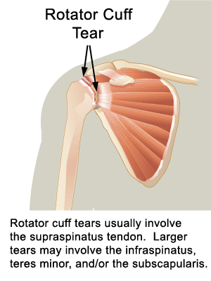
Alterations in function after rotator cuff tears in an animal model

Perry, S. M.; Getz, C. L.; Soslowsky, L. J.
J Shoulder Elbow Surg, Nº 18(2): 296-304

ABSTRACT

Hypothesis: This study utilized an animal model of multiple rotator cuff tendon tears to investigate alterations in the remaining (intact) rotator cuff tendon mechanical properties.

Materials and Methods: 24 rats served as uninjured controls, while 72 rats were divided among the tendon detachment groups (supraspinatus tendon detachment, supraspinatus+infraspinatus tendon detachment, supraspinatus+subscapularis tendon detachment). Mechanical testing was performed at 4 and 8 weeks post-injury.


Results: The remaining (intact) rotator cuff tendons had decreased mechanical properties in the presence of rotator cuff tears. Remaining (intact) subscapularis and infraspinatus tendon cross-sectional area increased, while tendon modulus decreased after both one and two tendon tears. Additionally, the remaining (intact) tendon cross-sectional areas continued to increase with time post-injury.

Discussion: This is the first animal study to examine the normal rotator cuff tendons and the remaining (intact) rotator cuff tendons after tendon detachment or tears.


We found that that the remaining (intact) rotator cuff tendons have altered mechanical properties with different tear sizes. These alterations could potentially lead to further tendon damage and tear progression

Conclusion: Results demonstrate not only differences in the uninjured rotator cuff tendons, but also that tears of one or two tendons affect the remaining (intact) tendons.


Keywords: Animal model; biomechanics; supraspinatus tendon; infraspinatus tendon; subscapularis tendon; shoulder; rotator cuff; tendon injury

Level of evidence: Level 1; Controlled laboratory study.

ARTIGO DO MÊS - (19/2009)


Prospective analysis of arthroscopic rotator cuff repair: Subgroup analysis
Nho, S. J.; Shindle, M. K.; Adler, R. S.; Warren, R. F.; Altchek, D. W.; Macgillivray, J. D.
J Shoulder Elbow Surg. 2009 Mar 6. [Epub ahead of print]

http://www.ncbi.nlm.nih.gov/pubmed/19269861?dopt=Citation

ABSTRACT

Background: The rotator cuff registry was established to prospectively evaluate the effectiveness of arthroscopic rotator cuff repair. The purpose of the present study is to report the preliminary data at the one-year and two-year time point, and perform subgroup analysis to identify factors that may affect outcome.

Methods: A total of one hundred ninety-three patients underwent all-arthroscopic repair of a rotator cuff tear and met the inclusion criteria, and 127 (65.8%) completed two-year follow-up. The outcome measurements included physical examination, manual muscle testing, the American Shoulder and Elbow Surgeons (ASES) score and ultrasonography.
Results: The pre-operative ASES score was 52.37 and improved to 83.88 at one year and 92.65 at two years. The percent healing for all patientswas 64.10% at 3 months and 64.34% at 1 year. At two years, there was a significant increase in the percentage of healed tendon at 75.42%compared to the three-month and the one-year timepoints.
Patients with intact tendons had an ASES score of 93.9 compared to tendon defects with a score of 88.0. Gender, tear size, and acromioclavicular joint involvement have a significant effect on ASES score.

Conclusion: Rotator cuff characteristics such as tear size, biceps pathology, acromioclavicular joint pathology, and tissue quality have a significant effect on post-operative tendon integrity. Arthroscopic rotator cuff repair demonstrates significant improvement in clinical outcomes and good rate of healing by post-operative ultrasound. Longer term studies are necessary to determine the efficacy over time.
Keywords: Arthroscopic rotator cuff repair; healing; ultrasound
Level of evidence: 3; Retrospective cohort study.
2009 Journal of Shoulder and Elbow Surgery Board of Trustees.


(...) The degree of rotator cuff size and associated pathology appear to have a dramatic affect on postoperative tendon integrity as well as age, to a lesser extent (...)

terça-feira, 19 de maio de 2009

LESÕES DE SOBRECARGA NO OMBRO DOS TENISTAS (4)

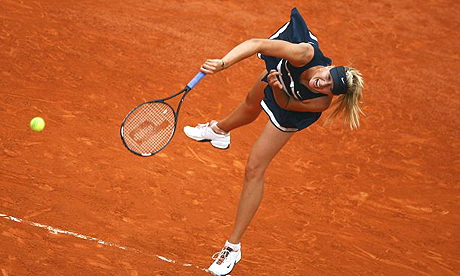
No dia 28 de Janeiro de 2009 escrevíamos um post sobre LESÕES DE SOBRECARGA NO OMBRO DOS TENISTAS (1) a propósito da lesão do ombro de Maria Sharapova (ex nº 1 do mundo).

Relembremos o que citámos na altura "M. Sharapova will pull out of next month’s Summer Olympics in Beijing, China. Sharapova withdrew from the Rogers Cup Wednesday evening, shortly after completing her second round match against Marta Domachowska. “After yesterday’s match I knew there was something seriously wrong with my shoulder,” wrote Sharapova on her official website.“After taking a few different exams and MRI’s this morning, the doctors found two small tears in the tendons of my shoulder.

Sabemos que acabou por ser operada ao ombro - Lesão parcial da coifa dos rotadores - depois de tentar tratamento não cirúrgico e que todo o processo de recuperação da lesão inicial afastou-a das competições praticamente 9 meses.

Competiu ao mais alto nível agora, no Torneio de Varsóvia (18 de Maio de 2009). Foi amplamente noticiado como in blog Bola amarela: "O dia foi de festa para o circuito feminino que hoje viu o regresso de uma das suas mais acarinhadas campeãs. Em Varsóvia, Maria Sharapova regressou após 9 meses longe dos courts e venceu a italiana Tathiana Garbin.

"No cômputo geral as ilações tiradas do regresso de Maria são bastante positivas, pese embora alguns dados menos entusiasmantes como a natural falta de confiança e o baixo ritmo no seu gesto de serviço. O saque foi mesmo um dos principais problemas da russa que cometeu 9 duplas-faltas, duas delas em match-points, situação que certamente terá estreita ligação com a lesão no ombro que a retirou de competição durante meses.

Volto a reforçar o que então escrevi em 3 post sobre as lesões de sobrecarga no ombro dos tenistas no final de Janeiro/2009 e que também é comum noutros atletas onde o gesto de lançamento/remate seja muito frequente/exigente (voleibol, andebol, lançamento do dardo, pólo aquático).

As exigências físicas colocadas ao ombro dos tenistas em termos de coordenação neuro-motora, amplitudes de movimentos solicitadas, cargas impostas e picos de velocidades (aceleração/desaceleração) são particularmente elevadas em determinados gestos como o serviço e o smash.

Uma última lição para muitos atletas, treinadores, médicos e fisioterapeutas: Não há tratamentos milagrosos e algumas das lesões exigem tempo, respeito pelos tempos de regeneração, abordagem multidiciplinar e trabalho.

Raul Oliveira, Fisioterapeuta

Equilibri_us - Gabinete de Fisioterapia

Av. D. João I, nº 8, Oeiras

309 984 508 / 917231718
raulov@netcabo.pt

Faculdade de Motricidade Humana
domingo, 17 de maio de 2009

INSTABILIDADE CRÓNICA DA TIBIO-TÁRSICA (3)

Tipos de medidas/factores sensório-motores relacionados com a instabilidade crónica da tibio-társica:

1) Sentido de posição e movimento articular - factor proprioceptivo

2) Excitabilidade e padrão de recrutamento neuromuscular

3) Mecanismos de pré-antecipação e/ou de ajustamento postural

4) Mecanismos reflexos de origem artrocinemática: depende da eficiência e qualidade da informação detectada em 1) e dos processos desenvolvidos em 2) e 3)

5) Força e coordenação neuromuscular

6) Controle postural dinâmico e equilíbrio eficiente em situações instáveis e/ou imprevistas: interacção entre sistemas perceptivos e sistemas de acção


7) Mecânica e fisiologia da marcha, corrida, saltos e gestos desportivos sobretudo nas fases de recepção (landing activities) e desaceleração.


Qualquer programa de intervenção da Fisioterapia neste tipo de condições deve incluir e desenvolver este tipo factores, depois de despistar lesões associadas (ver LESÕES CAPSULO-LIGAMENTARES DO TORNOZELO (4) e LESÕES CAPSULO-LIGAMENTARES DO TORNOZELO (4)

Raul Oliveira, Fisioterapeuta
Equilibri_us - Gabinete de Fisioterapia
Av. D. João I, nº 8, Oeiras
309 984 508 /917231718
raulov@netcabo.pt
Faculdade de Motricidade Humana

quarta-feira, 13 de maio de 2009

ARTIGO DO MÊS - (18/2009)


Treatment of common deficits associated with chronic ankle instability
Holmes, A.; Delahunt, E. Sports Med, 2009 (3):207-24
http://www.ncbi.nlm.nih.gov/pubmed/19290676?dopt=Citation


ABSTRACT:

Lateral ankle sprains are amongst the most common injuries incurred by athletes, with the high rate of reoccurrence after initial injury becoming of great concern.

Chronic ankle instability (CAI) refers to the development of repetitive ankle sprains and persistent residual symptoms post-injury. Some of the initial symptoms that occur in acute sprains may persist for at least 6 months post-injury in the absence of recurrent sprains, despite the athlete having returned to full functional activity.

CAI is generally thought to be caused by mechanical instability (MI) or functional instability (FI), or both. Although previously discussed as separate entities, recent research has demonstrated that deficits associated with both MI and FI may co-exist to result in CAI.

For clinicians, the main deficits associated with CAI include deficits in proprioception, neuromuscular control, strength and postural control.

Based on the literature reviewed, it does seem that subjects with CAI have a deficit in frontal plane ankle joint positional sense. Subjects with CAI do not appear to exhibit any increased latency in the peroneal muscles in response to an external perturbation.

Preliminary data suggest that feedforward neuromuscular control may be more important than feedback neuromuscular control and interventions are now required to address deficits in feed-forward neuromuscular control.

Balance training protocols have consistently been shown to improve postural stability in subjects with CAI. Subjects with CAI do not experience decreased peroneus longus strength, but instead may experience strength deficits in the ankle joint invertor muscles.

These findings are of great clinical significance in terms of understanding the mechanisms and deficits associated with CAI. An appreciation of these is vital to allow clinicians to develop effective prevention and treatment programmes in relation to CAI.

terça-feira, 12 de maio de 2009

INSTABILIDADE CRÓNICA DA TIBIO-TÁRSICA (2)

A lesão capsulo-ligamentar da tíbio-társica, resultante normalmente de mecanismos de entorse, é das lesões mais comuns, e após dois ou mais entorses de média gravidade num reduzido período de tempo, pode surgir a instabilidade crónica do tornozelo que, por sua vez, aumenta o risco de novos entorses (Ross, et al., 2008), promovendo a incapacidade funcional nas actividades mais exigentes.

Recordemos que a instabilidade crónica da tíbio-társica pode ser considerada uma disfunção associada a 2 factores potenciais que interagem: a instabilidade mecânica passiva e a instabilidade dinâmica funcional.

ver INSTABILIDADE CRÓNICA DA TIBIO-TÁRSICA (1)


A instabilidade dinâmica funcional está relacionada com perturbações cinestésicas que alteram a actividade reflexa artrocinemática após lesão ligamentar (Hertel, 2002, Claire E. et al, 2004), com a repetição de mecanismos de entorse por ineficiência desses mecanismos de protecção/estabilidade articular e ainda com a sensação subjectiva de instabilidade (Karlsson & Chan, 2005).

Sempre que há uma lesão capsulo-ligamentar há também uma perturbação da informação proveniente dos mecanoreceptores articulares, causando desaferenciação parcial da articulação. Este facto promove altera o normal funcionamento do controlo neuromuscular, resultando numa diminuição da estabilização da articulação, por alteração dos padrões neuromotores, e por último, contribuindo para mecanismos de entorse de repetição cada vez mais fáceis de acontecer originando perturbações na funcionalidade do segmento (Lephart & Fu, 2002).

Como a informação proprioceptiva é uma das fontes de informação envolvidas no controle da postura e do equilíbrio dinâmico, as lesões capsulo-ligamentares afectam negativamente esse controle, em virtude da laxidão ligamentar residual e dos défices sensório-motores que surgem sobretudo se o tratamento não for o mais adequado.

Nos quadros de instabilidade crónica da tibio-társica, têm sido referidas modificações na percepção de posição da articulação (Chang et al., 2001), alterações no tempo de reacção dos peroniais ao movimento de inversão/flexão plantar repentino e alterações da velocidade de condução do impulso nervoso (Lephart & Fu, 2000)


O binómio funcional mobilidade/estabilidade do tornozelo, como em qualquer outra articulação, depende da interacção entre factores de natureza estática (carga, morfologia osteo-articular, estruturas capsulo-ligamentares e forças de coesão intra-articular) e factores dinâmicos ou funcionais como os relacionados com a função neuromuscular (força, stiffness e coordenação intra e intermuscular) assim como a operacionalidade/eficiência do controlo motor assegurado pelo sistema nervoso.


Freeman et al. (1965) afirmaram que uma lesão da tíbio-társica pode danificar a função dos receptores aferentes localizados nos ligamentos e cápsula articular, levando a uma alteração do sistema de controlo postural.
As alterações da propriocepção articular, após a ocorrência de lesão cápsulo-ligamentar da articulação tíbio-társica, diminuem o feedback sensorial aferente da articulação lesionada, resultando num aumento da oscilação postural (Lephart et al., 1997). Estes défices proprioceptivos dos ligamentos do tornozelo podem também ocorrer devido a fraqueza dos músculos responsáveis pela estabilização e oscilação postural (Lephart & Fu 2000).
Raul Oliveira, Fisioterapeuta
Equilibri_us - Gabinete de Fisioterapia
Av. D. João I, nº 8, Oeiras
309 984 508 /917231718
Faculdade de Motricidade Humana